- The brain with metastasis
Endothelial cells
- Do metastases require the generation of new vessels to thrive?

Vascular co-option
García-Gómez P and Valiente M. (2020). Tumor vascularization. Elsevier.
Vascular co-option in brain metastasis.
García-Gómez P and Valiente M. Angiogenesis. (2020).
Serpins promote cancer cell survival and vascular co-option in brain metastasis.
Valiente M et al. Cell. (2014).
Pericyte-like spreading by disseminated cancer cells activates YAP and MRTF for metastatic colonization.
Er E et al. Nature Cell Biology. (2018).
Protocol to generate murine organotypic brain cultures for drug screening and evaluation of anti-metastatic efficacy
Lucía Zhu, Lauritz Miarka, Patricia Baena, María Perea-García, Manuel Valiente. STAR Protocols (2023)
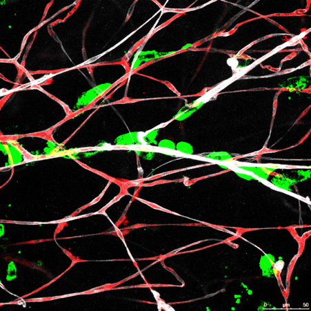
In the brain (and elsewhere) the initiation of metastasis relies on the ability of cancer cells to interact with pre-existing vessels. This perivascular niche is exploited by metastasis initiating cells to favor organ colonization in a process termed vascular co-option. Why is vascular co-option required for the initiation of metastasis is unknown and which are the molecular mediators of such process poorly described.
We extensively used organotypic brain cultures to study the process of vascular co-option both to understand the influence of the physical interaction on co-opting cancer cells as well as on co-opted endothelial cells. Such a unique but also reproducible phenomena among models of brain metastasis is being dissected ultrastructurally with electron microscopy as well as interrogated with unprecedented detail at the molecular level. We have expanded the research tools available to study vascular co-option by using multicolor reporter time-lapse video-microscopy, specific AAVs targeting endothelial cells as well as GEMM that could help us to target specific genes in this compartment of the microenvironment.
We envision that exploiting vascular co-option is a unique opportunity to develop innovative strategie to prevent metastasis from happening.